


RENAL STONES
Aetiopathogenesis
1. Infection: Organisms such as Proteus, Pseudomonas, Klebsiella produce recurrent UTI. These organisms produce urea, cause stasis of urine and precipitate stone formation. Nucleus of the stone may harbour these bacteriae.
2. Hot climates cause increase in concentration of solutes, resulting in precipitation of calcium and formation of calcium oxalate stones.
4. Metabolic causes • Hyperparathyroidism increases serum calcium levels resulting in hypercalcinosis and pelvic stones. • Gout increases uric acid levels and causes multiple uric acid stones.
5. Immobilisation: Paraplegic patients secrete large amounts of calcium in the urine resulting in calcium oxalate stones (they pass skeletons in urine).
6. Decreased urinary citrate: Citric acid (300-900 mg/24 hours) keeps the urinary pH low. When citric acid levels decrease, it promotes precipitation of urinary calcium. Citrate excretion is under hormonal control.
7. Inadequate urinary drainage as in cases of horseshoe kidney, unascended kidneys are more vulnerable for development of stones due to stasis.
8. Randall's plaques: Randall has suggested that initially a small erosion or an ulcer develops at the tip of renal papilla on which minute concretions or minor calcium particles get deposited and give rise to stone formation.
Types of renal stones
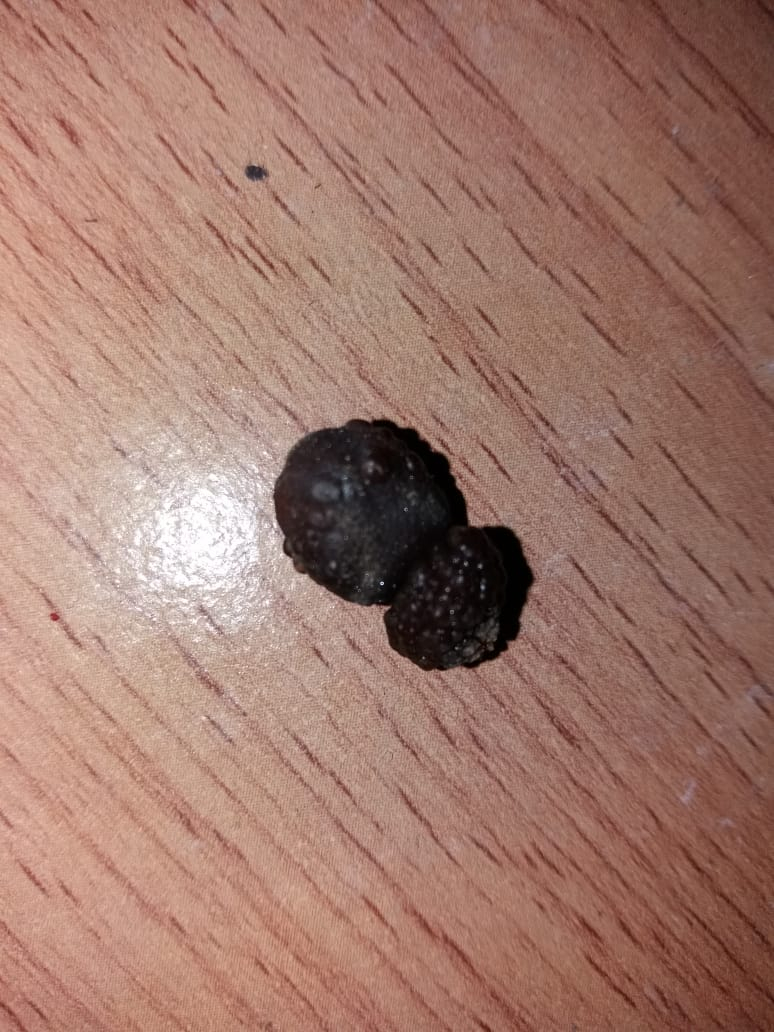
1. Calcium oxalate stone
• Called as mulberry calculi
• Common type of stone
• It is irregular having sharp projections
• Oxalate stone is hard and single
• Produces haematuria very early, resulting in deposition of blood over the stone giving a dark colour to the stone.
• It occurs in infected urine
• Contains alternate layer of calcium and bacterial vegetation. It is visualised in plain X-ray KUB.
2. Uric acid stone
• Multiple, small, hexagonal, faceted, yellow coloured.
• Contain calcium oxalate which makes them opaque. Pure uric acid stones are radiolucent.
• Occur in acidic urine
• Common in patients who consume red meat
• Best responsive to lithotripsy
3. Phosphate stone
• Smooth, round
• Consists of triple phosphate of calcium, magnesium and ammonium.
• Dirty white to yellow in colour
• Commonly occur in renal pelvis and tend to grow in alkaline urine.
• As it enlarges in the pelvis, it grows within the major and minor calyces and slowly forms staghorn calculus. This calculus produces recurrent urinary tract infection and haematuria and slowly damages the renal parenchyma
4. Cystine calculus
• Cystinuria is an inborn error of metabolism which occur due to decreased resorption of cystine from the renal tubules.
• Occurs in young girls at puberty
• Increased excretion of cystine in urine results in cystine calculus.
• Stones are hard and radio-opaque due to sulphur.
Clinical features
• Renal pain: Dull aching to pricking type of pain posteriorly in the renal angle formed by the sacrospinalis and 12th rib. Murphy's kidney punch test demonstrates tenderness at renal angle. The same pain may some times be felt anteriorly in the costal margin. Hence, it is described as costovertebral pain. Nausea and vomiting is due to intense sympathetic stimulation caused by stretching of renal capsule mediated by coeliac plexus.
• Ureteric colic: When the stone is impacted in the pelviureteric junction or anywhere in the ureter, it results in severe colicky pain originating at the loin and radiating to the groin, testicles, vulva and medial side of the thigh. This may be associated with strangury. The referred pain is due to irritation of the genitofemoral nerve.
• Haematuria is common with renal stone because the majority of stones are oxalate stones. The quantity of blood lost is small but it is fresh blood.
• Recurrent UTI: Fever with chills and rigors, burning micturition, pyuria may occur, along with increased frequency of micturition.
• Guarding and rigidity of the back and abdominal muscles during severe attack of pain.
Complications
1. Calculous hydronephrosis occurs due to back pressure producing renal enlargement. Stretching of the renal capsule results in pain. In such cases, an associated palpable kidney mass suggests hydronephrosis.
2. Calculous pyonephrosis: Infected hydronephrosis where in the kidney is converted into a bag of pus.
3. Renal failure: Bilateral staghorn stones may not be symptomatic until they present with uraemia and renal failure.
4. Squamous cell carcinoma: Long-standing stones increase the risk of carcinoma.
Investigations
1. Blood urea and creatinine to rule out renal failure.
2. Plain X-ray KUB
• To diagnose stones 90% of the renal stones are radioopaque.
• Enlarged renal shadow can be seen.
3. USG
• Presence of the stone can be diagnosis
• Exact size and location of the stone can be evaluated.
4. IVP
• To locate the stone exactly in relation to kidney and ureter and to assess renal function. A nonradiopaque stone can be seen as a filling defect. Hydronephrosis and hydronephroureterosis can also be seen.
• Now noncontrast CT scan and contrast CT scan are used for more accurate detection of causes of abdominal colic.
5. Urine for culture and sensitivity.
Nonoperative treatment
1. Conservative: Small stones less than 5 mm in size pass off with intake of copious amount of fluids and at times forced diuresis.
URETERIC STONE
Stones come down from pelvis of the kidney and may get impacted at any site of anatomical narrowing of ureter, namely:
1. Pelviureteric junction
2. Crossing of the iliac artery
3. Crossing of the vas deferens or broad ligament.
4. Site of entry into the bladder wall
5. Ureteric orifice
This may lead to hydroureteronephrosis, renal parenchymal atrophy, infection and pyonephrosis.
Clinical features
• Pain in the loin radiating to groin: Pain is severe, colicky, intolerable and lasts for a few hours. When stone descends into lower ureter, pain radiates to the testicles, labia majora and to the upper portion of thigh due to irritation of genitofemoral nerve. Colic lasts for about 4-6 hours and is relieved by antispasmodics, narcotics and NSAID.
• An attack of haematuria or pyuria
• Guarding and rigidity of the abdominal wall if present on the right side, is confused with acute appendicitis.
Investigations
Same as renal stone
Treatment
Most of the ureteric stones pass via naturalis (urine). The patient is asked to consume a lot of water and antispasmodics.
Prevention of stone disease
I. Metabolic work up of urine and blood for identifying metabolic causes. Example: Hyperparathyroidism to be ruled out by 24 hours urine analysis for calcium, phosphate and uric acid levels.
2. Fluid management: 1.5 L/day
3. Dietary adjustments: Red meat to be avoided (rich in uric acid)
4. Drug treatment
• Xyloric, sodium bicarbonate: Uric acid stones
• Potassium citrate: Calcium stones
• Thiazides small dose: Calcium stones
• D-penicillamine: Cystine stones
• Protease inhibitors: Infection stones against E. coli
5. Ultrasound to be done once in 6 months.
TREATMENT AT DR. SOHAN LAL CLINIC
The integrated POLYCLINIC facility offers patients to select their treatment either from the Department of Homeopathy or from the Department of Medicine.
We provide scientific, research-based, and professional services to people across the world, aiming to achieve the highest success rate.

A clinic of national fame with 92 Years of legacy in healthcare. Dr. Sohan Lal Clinic is a state-of-the-art Polyclinic offering integrated healthcare facilities, including online consultation and online medicine delivery. We are an established BRAND- highly trusted for having a high success rate in treatment and being globally affordable.
